Beans + Sprouts + Seeds
Lectin from green speckled lentil seeds (Lens culinaris) triggered apoptosis in nasopharyngeal carcinoma cell lines
Yau Sang Chan1, Huimin Yu1, Lixin Xia1 and Tzi Bun Ng2*
Abstract
Background
The green speckled lentil seed (Lens culinaris) lectin (GSLL) exhibits hemagglutinating activity, and possesses some properties distinct from those of other lentil lectins (e.g., molecular size, biological activities) that deserve further investigation. This study aims to investigate the basic properties (e.g., molecular size, amino acid sequence, sugar specificity) and biological activities (e.g., antiproliferative activity) of GSLL.
Methods
GSLL was purified by successive fractionation on SP-Sepharose, Affi-gel blue gel, Mono Q, and Superdex 75. The biochemical properties of GSLL were investigated by SDS-PAGE, mass spectrometry, N-terminal amino acid sequencing, and sugar inhibition tests. For the biological activities, purified lyophilized GSLL was sterilized, adjusted to concentrations from 1 to 0 mg/mL (by twofold serial dilution) in Dulbecco’s modified Eagle’s medium with fetal bovine serum, and examined by using the MTT assay, flow cytometry, and western blotting after treatment of nasopharyngeal carcinoma CNE1 and CNE2 cell lines with the lectin.
Results
GSLL appeared as a 21-kDa band in non-reducing SDS-PAGE. It was composed of two subunits with molecular sizes of 17 and ~4 kDa. It exhibited specificity in binding to glucose and mannose, as well as glucosides and mannosides. Mass spectrometry and N-terminal amino acid sequencing revealed similarity of GSLL to L. culinaris lectin (LcL), especially higher coverage of the β-chain of LcL. A 48-h treatment with GSLL exerted antiproliferative effects on nasopharyngeal carcinoma CNE1 and CNE2 cell lines with significant inhibition at 0.125 mg/mL (P < 0.001) and 1 mg/mL (P = 0.004), respectively, and these effects were attenuated in the presence of glucose and mannose. GSLL induced apoptosis in nasopharyngeal carcinoma CNE1 cells, with detectable phosphatidylserine externalization, mitochondrial depolarization, and cell cycle arrest. Western blot analysis suggested that GSLL triggered the extrinsic apoptotic pathway involving caspase 3, 8, and 9 in CNE1 cells.
Conclusion
GSLL possessed some different properties from LcL (e.g., lower pI), and increased caspase 3, 8, and 9 activity in CNE1 cells.
Source : Chinese Medicine Journal
Link to Full Article
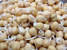
Isoflavones Extracted from Chickpea Cicer arietinum L. Sprouts Induce Mitochondria-Dependent Apoptosis in Human Breast Cancer Cells
- Hua Chen1,2,3,
- Hai-Rong Ma1,2,*,
- Yan-Hua Gao1,2,
- Xue Zhang4,
- Madina Habasi1,2,
- Rui Hu1,2,3 and
- Haji Akber Aisa1,2,*
Abstract
Isoflavones are important chemical components of the seeds and sprouts of chickpeas. We systematically investigated the effects of isoflavones extracted from chickpea sprouts (ICS) on the human breast cancer cell lines SKBr3 and Michigan Cancer Foundation-7 (MCF-7). 3-(4,5-dimethylthiazol-2-yl)-2,5-diphenyltetrazolium bromide assays showed that ICS (10–60 µg/mL) significantly inhibited the proliferation of both cell lines in a time-dependent and dose-dependent fashion. Wright-Giemsa staining as well as annexin V-fluorescein isothiocyanate and propidium iodide (Annexin V/PI) staining showed that ICS significantly increased cytoclasis and apoptotic body formation. Quantitative Annexin V/PI assays further showed that the number of apoptotic cells increased in a dose-dependent manner following ICS treatment. Semiquantitative reverse transcription PCR showed that ICS increased the expression of the apoptosis-promoting gene Bcl-2-associated X protein and decreased the expression of the antiapoptotic gene Bcl-2. Western blot analysis showed that treatment of SKBr3 and MCF-7 cells with ICS increased the expression of caspase 7, caspase 9, P53, and P21 in a dose-dependent manner. Flow cytometry assays using the fluorescent probe 3,3′-dihexyloxacarbocyanine iodide showed a dose-dependent decrease in mitochondrial membrane potential following ICS treatment. Treatment using ICS also induced a dose-dependent increase in reactive oxygen species production. This is the first study to demonstrate that ICS may be a chemopreventive or therapeutic agent against breast cancer.
Source : Phytotherapy Research
Link to Full Article