Bangladeshi Medicinal Plant Extracts
Cytotoxicity screening of Bangladeshi medicinal plant extracts on pancreatic cancer cells
Sherine George1, Siddharth V. Bhalerao2, Erich A. Lidstone1, Irfan S. Ahmad3,4, Atiya Abbasi5, Brian T. Cunningham1,2,3, Kenneth L. Watkin3,6*
1Department of Bioengineering, University of Illinois at Urbana Champaign, USA
2Department of Electrical and Computer Engineering, University of Illinois at Urbana Champaign, USA
3Center for Nanoscale Science and Technology, Micro and Nanotechnology Laboratory, University of Illinois at Urbana Champaign, USA
4Agricultural and Biological Engineering, University of Illinois at Urbana Champaign, USA
5International Center for Chemical and Biological Sciences, University of Karachi, Pakistan
6Beckman Institute for Advanced Science and Technology, Bio-Imaging Science and Technology Group, University of Illinois at Urbana Champaign, USA
Background
Pancreatic cancer is the fourth leading cause of cancer-related death in both sexes in the United States [1]. Although Gemcitabine is the current first-line chemotherapeutic administered for metastatic pancreatic cancer, this line of treatment has been met with limited survival and symptomatic outcomes [2, 3] resulting in research interest in exploring new alternatives for treatment and prevention. Natural products play a dominant role in the discovery of such new drugs, as over 60% of approved drugs or those in late stages of development (during 1989-1995) are of natural origin [4]. Examples of clinically useful antitumor agents derived from plants
include paclitaxel, vincristine, and camptothecin. Many of these plant-derived anticancer agents have been discovered through large-scale screening programs [5]. Furthermore, the broadreaching support and continuation of studies of plant extracts with implications in pancreatic cancer treatment are indicative of the continued role that natural products play in the drug discovery process [6, 7].
This study provides data on the cytotoxic potential of 56 extracts derived from 44 different plants used in Bangladeshi folk medicine. A three-tiered screening system was designed, in which all extracts were first screened for their ability to induce death in the Panc-1 cell line using a label-free photonic crystal (PC) biosensor assay. These experiments generated biosensor images of attached cells which were used to quantify cell proliferation changes in treated versus untreated cultures. Next, extracts that showed significant cytotoxicity to Panc-1 cells (>80% cell death at a testing concentration of 100 μg/mL) in the PC biosensor assay were
tested using a colorimetric MTT assay on two additional pancreatic cell lines (Mia-Paca2, and Capan-1). Toxicity to a normal foreskin Hs68 fibroblast cell line was studied as a control Finally, the extract showing the highest cytotoxicity in all three cancer cell lines was evaluated for its apoptotic activity via a caspase-3 quantification assay.
Methods:
56 extracts of 44 unique medicinal plants were studied. The extracts were screened for cytotoxicity against the pancreatic adenocarcinoma cell line Panc-1, using a label-free biosensor assay. The top cytotoxic extracts identified in this screen were tested on two additional pancreatic cancer cell lines (Mia-Paca2 and Capan-1) and a fibroblast cell line (Hs68) using an MTT proliferation assay. Finally, one of the most promising extracts was studied using a caspase-3 colorimetric assay to identify induction of apoptosis.
Results:
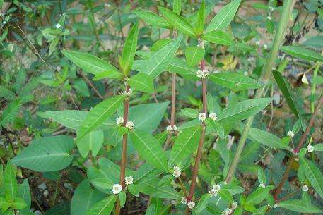
Crude extracts of Petunia punctata, Alternanthera sessilis, and Amoora chittagonga showed cytotoxicity to three cancer cell lines with IC50 values ranging between 20.3 – 31.4 μg/mL, 13.08 – 34.9 μg/mL, and 42.8 – 49.8 μg/mL, respectively. Furthermore, treatment of Panc-1 cells with Petunia punctata was shown to increase caspase-3 activity, indicating that the observed cytotoxicity was mediated via apoptosis. Only Amoora chittagonga showed low cytotoxicity to fibroblast cells with an IC50 value > 100 μg/mL.
Conclusion:
Based upon the initial screening work reported here, further studies aimed at the identification of active components of these three extracts and the elucidation of their mechanisms as cancer therapeutics are warranted
Source : BMJ Complementary and Alternative Medicine
LINK TO FULL ARTICLE
Sherine George1, Siddharth V. Bhalerao2, Erich A. Lidstone1, Irfan S. Ahmad3,4, Atiya Abbasi5, Brian T. Cunningham1,2,3, Kenneth L. Watkin3,6*
1Department of Bioengineering, University of Illinois at Urbana Champaign, USA
2Department of Electrical and Computer Engineering, University of Illinois at Urbana Champaign, USA
3Center for Nanoscale Science and Technology, Micro and Nanotechnology Laboratory, University of Illinois at Urbana Champaign, USA
4Agricultural and Biological Engineering, University of Illinois at Urbana Champaign, USA
5International Center for Chemical and Biological Sciences, University of Karachi, Pakistan
6Beckman Institute for Advanced Science and Technology, Bio-Imaging Science and Technology Group, University of Illinois at Urbana Champaign, USA
Background
Pancreatic cancer is the fourth leading cause of cancer-related death in both sexes in the United States [1]. Although Gemcitabine is the current first-line chemotherapeutic administered for metastatic pancreatic cancer, this line of treatment has been met with limited survival and symptomatic outcomes [2, 3] resulting in research interest in exploring new alternatives for treatment and prevention. Natural products play a dominant role in the discovery of such new drugs, as over 60% of approved drugs or those in late stages of development (during 1989-1995) are of natural origin [4]. Examples of clinically useful antitumor agents derived from plants
include paclitaxel, vincristine, and camptothecin. Many of these plant-derived anticancer agents have been discovered through large-scale screening programs [5]. Furthermore, the broadreaching support and continuation of studies of plant extracts with implications in pancreatic cancer treatment are indicative of the continued role that natural products play in the drug discovery process [6, 7].
This study provides data on the cytotoxic potential of 56 extracts derived from 44 different plants used in Bangladeshi folk medicine. A three-tiered screening system was designed, in which all extracts were first screened for their ability to induce death in the Panc-1 cell line using a label-free photonic crystal (PC) biosensor assay. These experiments generated biosensor images of attached cells which were used to quantify cell proliferation changes in treated versus untreated cultures. Next, extracts that showed significant cytotoxicity to Panc-1 cells (>80% cell death at a testing concentration of 100 μg/mL) in the PC biosensor assay were
tested using a colorimetric MTT assay on two additional pancreatic cell lines (Mia-Paca2, and Capan-1). Toxicity to a normal foreskin Hs68 fibroblast cell line was studied as a control Finally, the extract showing the highest cytotoxicity in all three cancer cell lines was evaluated for its apoptotic activity via a caspase-3 quantification assay.
Methods:
56 extracts of 44 unique medicinal plants were studied. The extracts were screened for cytotoxicity against the pancreatic adenocarcinoma cell line Panc-1, using a label-free biosensor assay. The top cytotoxic extracts identified in this screen were tested on two additional pancreatic cancer cell lines (Mia-Paca2 and Capan-1) and a fibroblast cell line (Hs68) using an MTT proliferation assay. Finally, one of the most promising extracts was studied using a caspase-3 colorimetric assay to identify induction of apoptosis.
Results:
Crude extracts of Petunia punctata, Alternanthera sessilis, and Amoora chittagonga showed cytotoxicity to three cancer cell lines with IC50 values ranging between 20.3 – 31.4 μg/mL, 13.08 – 34.9 μg/mL, and 42.8 – 49.8 μg/mL, respectively. Furthermore, treatment of Panc-1 cells with Petunia punctata was shown to increase caspase-3 activity, indicating that the observed cytotoxicity was mediated via apoptosis. Only Amoora chittagonga showed low cytotoxicity to fibroblast cells with an IC50 value > 100 μg/mL.
Conclusion:
Based upon the initial screening work reported here, further studies aimed at the identification of active components of these three extracts and the elucidation of their mechanisms as cancer therapeutics are warranted
Source : BMJ Complementary and Alternative Medicine
LINK TO FULL ARTICLE